

Fluorescence and Micro-FTIR characteristics of typical liptinite at low temperature thermal conversion
-
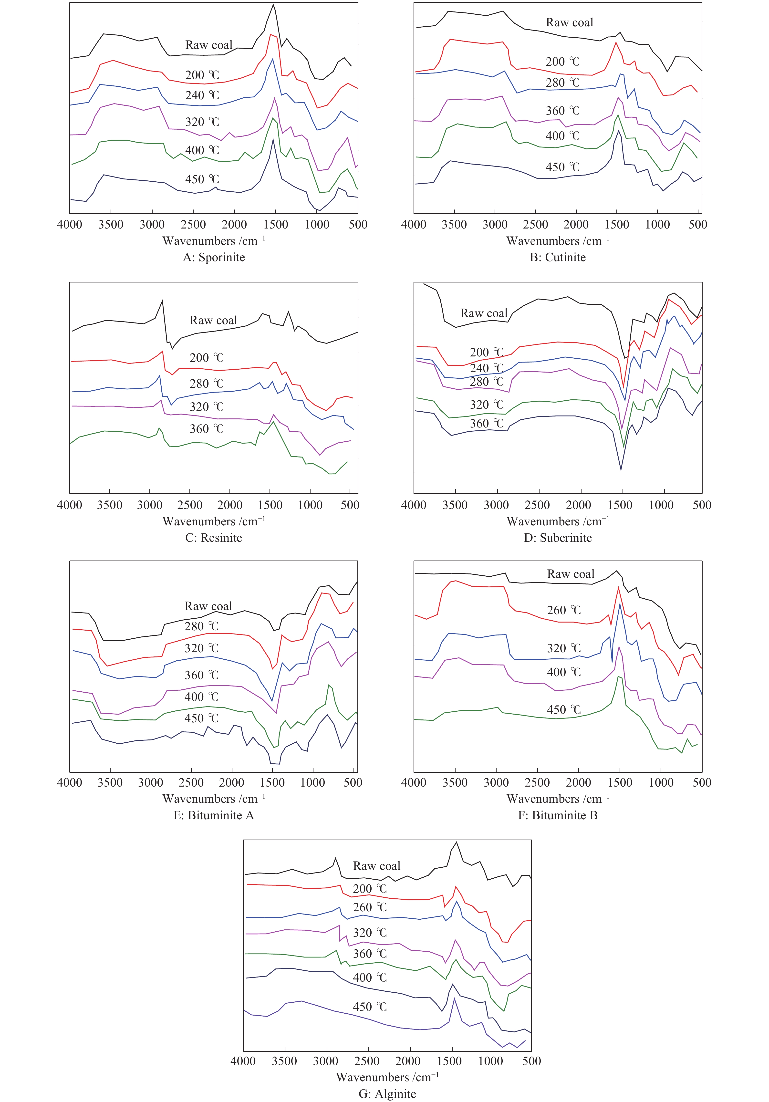
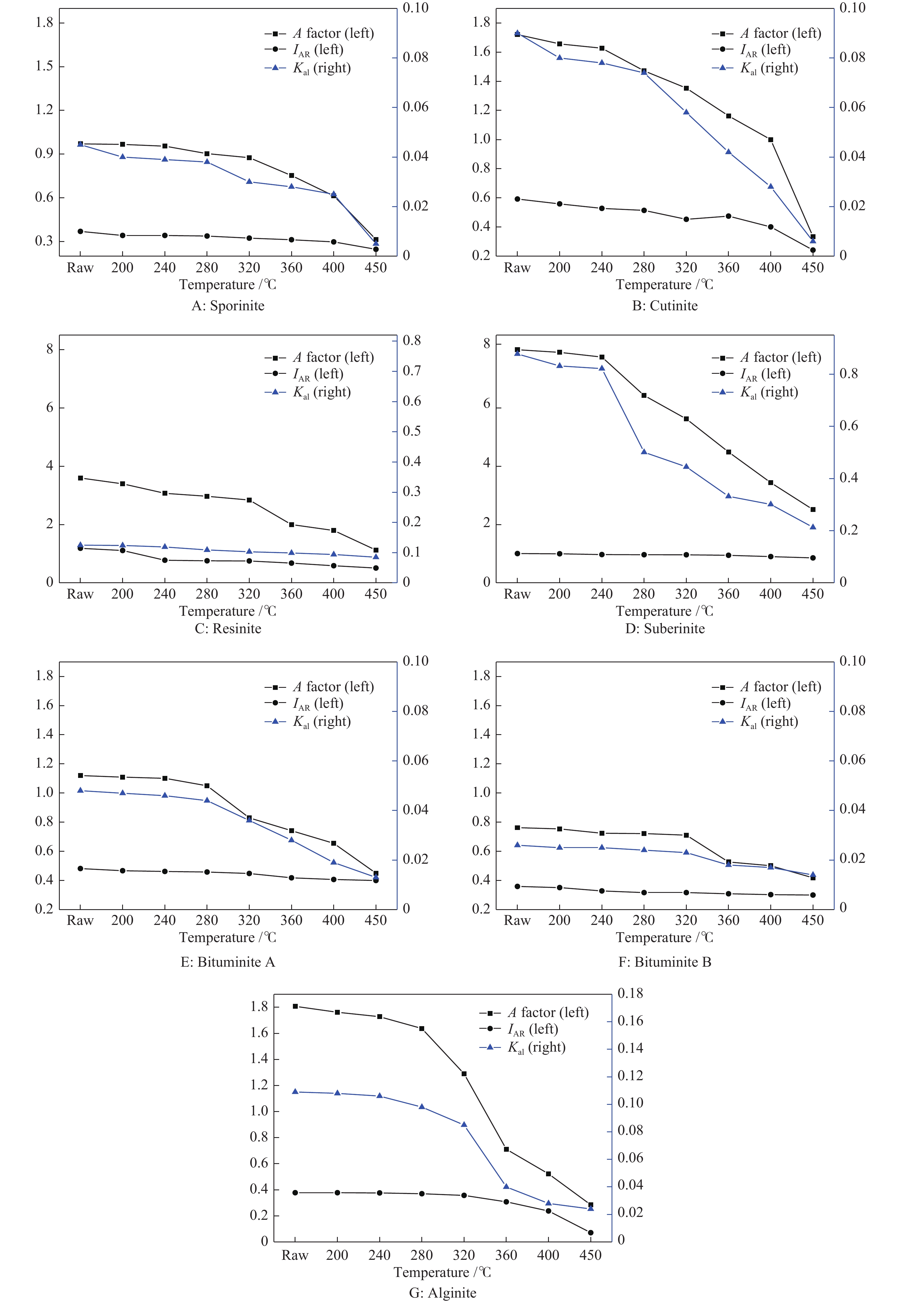
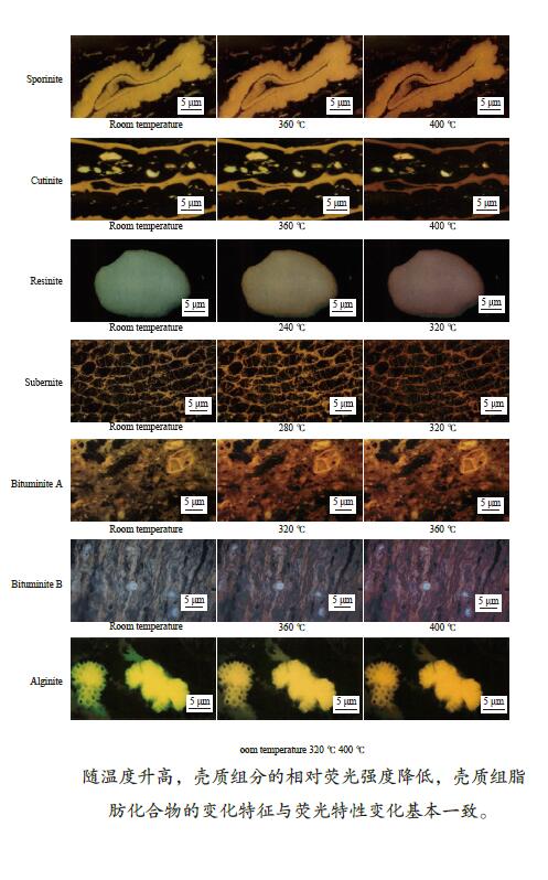
摘要: 煤中壳质组的种类及含量对热解焦油、煤气产率有重要的影响。综合利用显微镜热台、荧光显微分析和显微傅里叶红外光谱(Micro-FTIR)研究煤中典型壳质组的低温热转化( ≤ 450 ℃)特性。结果表明,随着温度升高,煤中壳质组分的相对荧光强度降低,最大荧光波长增大;树脂体和木栓质体的荧光特性在240 ℃开始变化,280−320 ℃变化显著;孢粉体、角质体和沥青质体A的荧光特性在280 ℃开始变化,320−360 ℃变化显著;藻类体的荧光特性从280 ℃开始变化,并持续到400 ℃;沥青质体B的荧光变化出现在320−360 ℃。藻类体的脂肪化合物的吸收峰最强,其次为沥青质体、树脂体、角质体和孢粉体;随着温度升高,壳质组的脂肪族和含氧基团逐渐减少,芳香烃含量相对增加。煤中壳质组分芳构化程度低,在低温热转化过程中基本保持不变;壳质组的富氢程度及脂肪化合物变化特征与荧光特性的变化基本一致。
-
关键词:
- 壳质组 /
- Micro-FTIR /
- 荧光特性 /
- 热解
Abstract: The subgroups and contents of liptinite have significant influence on yields of tar and gas in pyrolysis.The heating stage microscope, fluorescence analysis and Micro-FTIR were used to study the characteristics of typical liptinite at low temperature thermal conversion. The results showed that the relative fluorescent intensities decreased and the maximum fluorescence wavelength increased as the pyrolysis temperature increase. The fluorescence characteristics of resinite and suberinite began to change at 240 ℃, and remarkable changed at 280−320 ℃. The fluorescence characteristics of sporinite, cutinite, and bituminite A began to change at 280 ℃, and remarkable changed at 320−360 ℃. The fluorescence characteristic of alginite began to change at 280 ℃, and lasting changed till 400 ℃. The fluorescence characteristic of bituminite B changed at 320−360 ℃. The absorption peak of aliphatic compound in alginite was strongest, and the peaks of bituminite, resinite, cutinite, and sporinite decreased in turn. The aliphatic compounds and oxygen-containing groups of liptinite group decreased and the content of aromatic hydrocarbon relatively increased as the temperature increase. The aromatization degree of liptinite group was low and basically remain unchanged at low temperature thermal conversion. The hydrogen-rich degrees and the change of aliphatic chains of liptinite group confirmed well with the fluorescence characteristics.-
Key words:
- liptinite /
- Micro-FTIR /
- fluorescence characteristic /
- pyrolysis
-
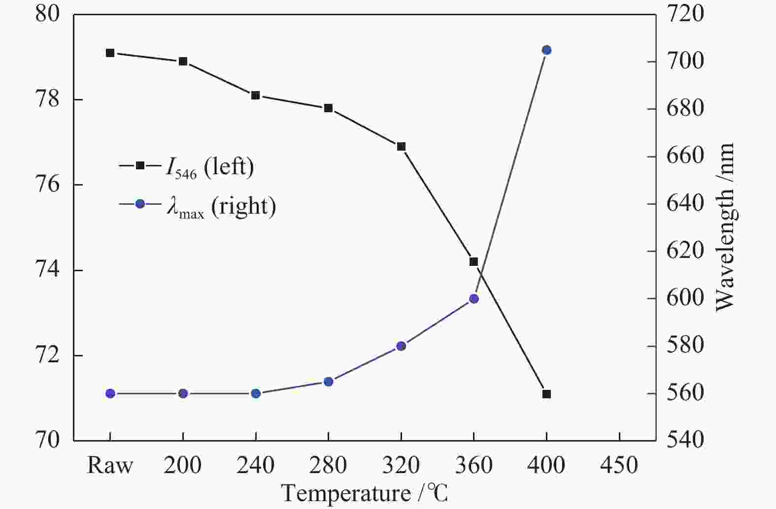
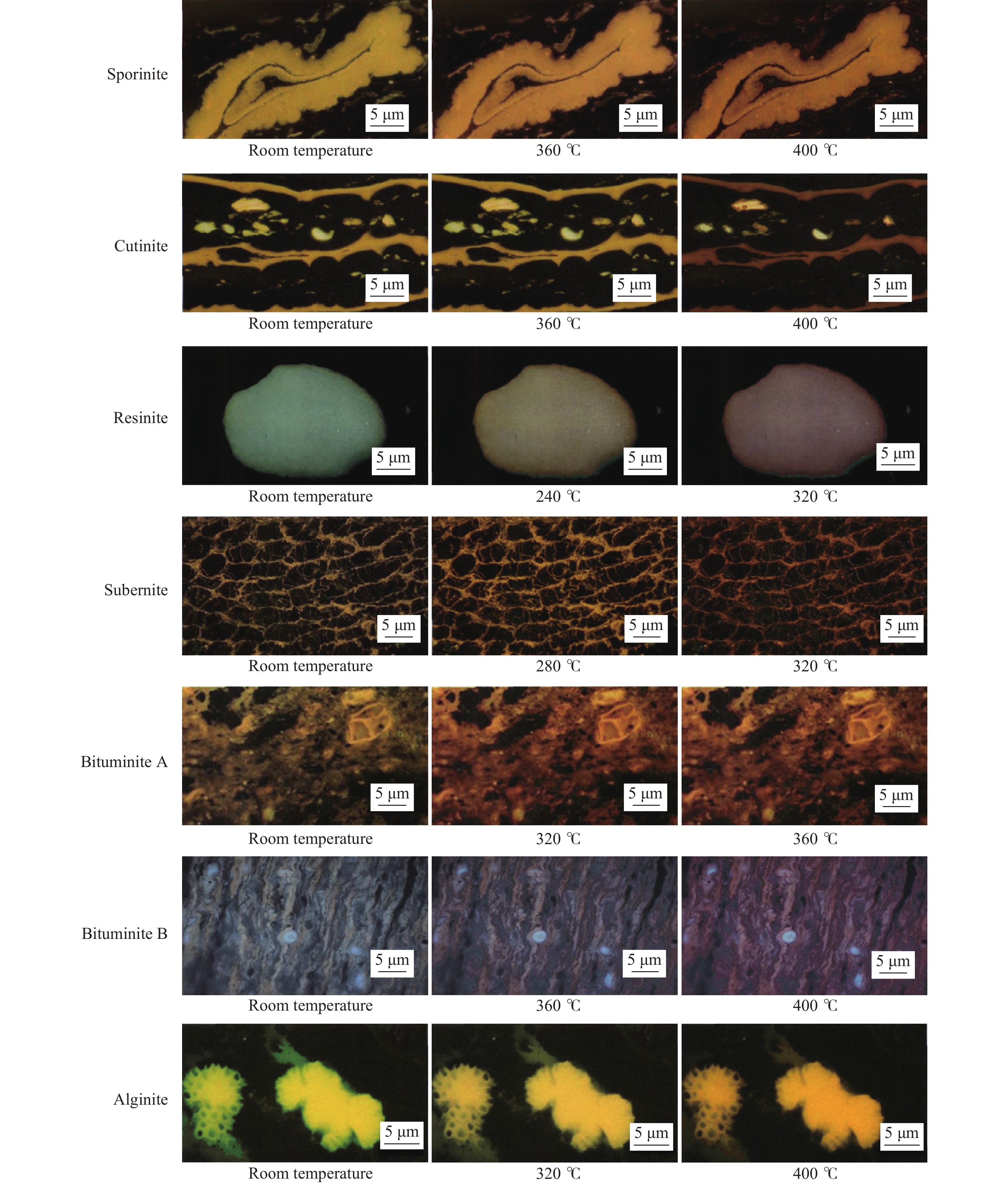
图 3 不同热转化温度下孢粉体的荧光特性
Figure 3 Fluorescence properties of sporinite at low temperature thermal conversion
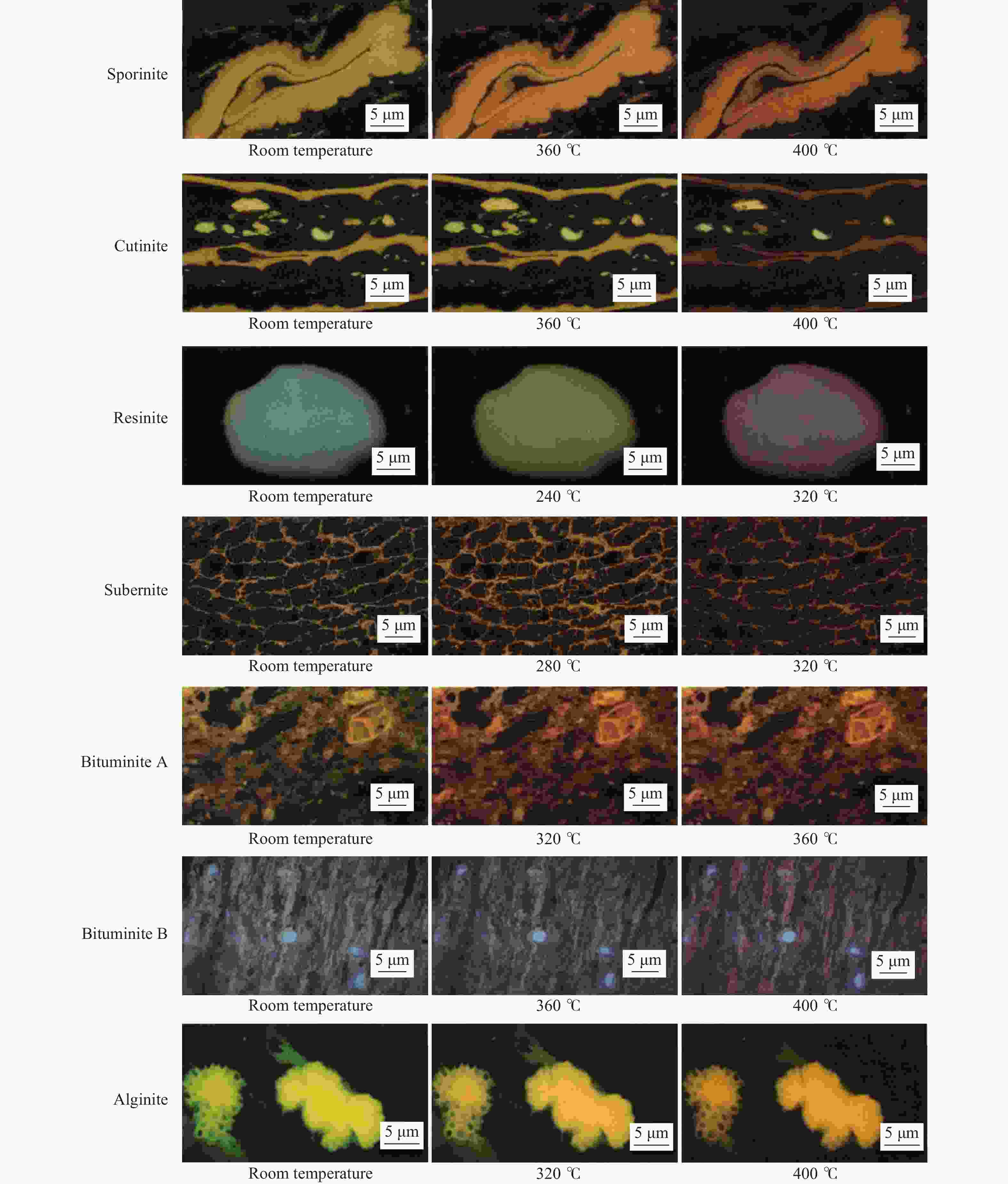
图 2 低温热转化过程中壳质组的荧光性质变化
Figure 2 Fluorescence properties of typical liptinite in low temperature thermal conversion
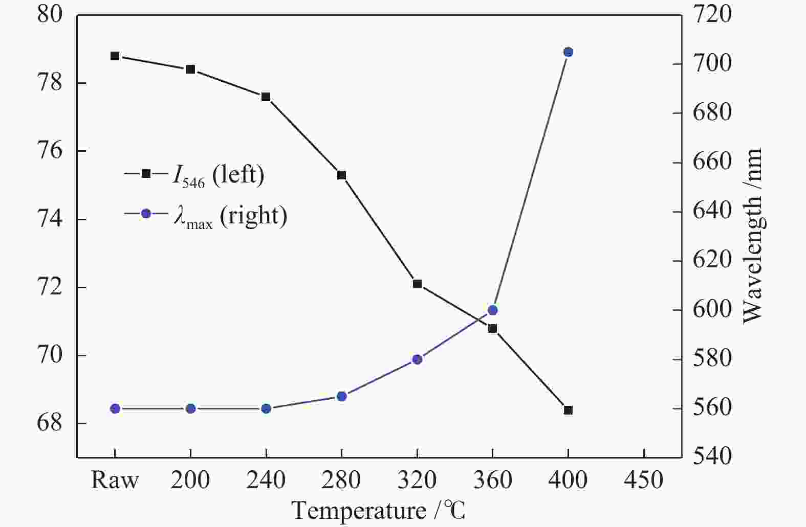
图 4 不同热转化温度下角质体的荧光特性
Figure 4 Fluorescence properties of cutinite at low temperature thermal conversion
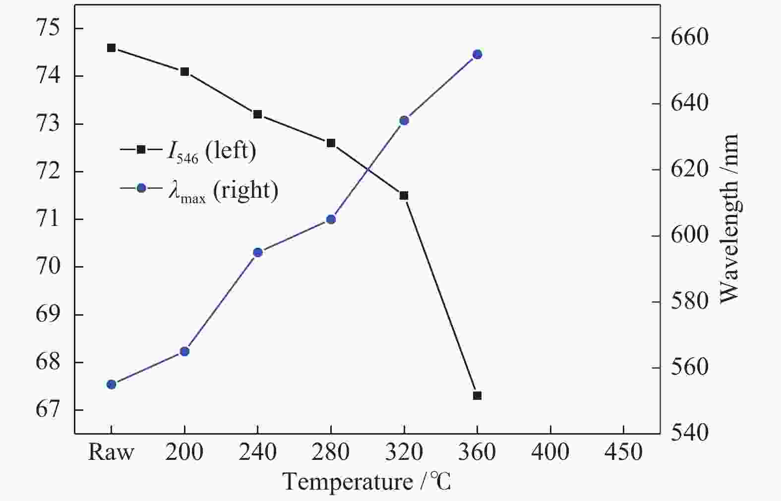
图 5 不同热转化温度下树脂体的荧光特性
Figure 5 Fluorescence properties of resinite at low temperature thermal conversion
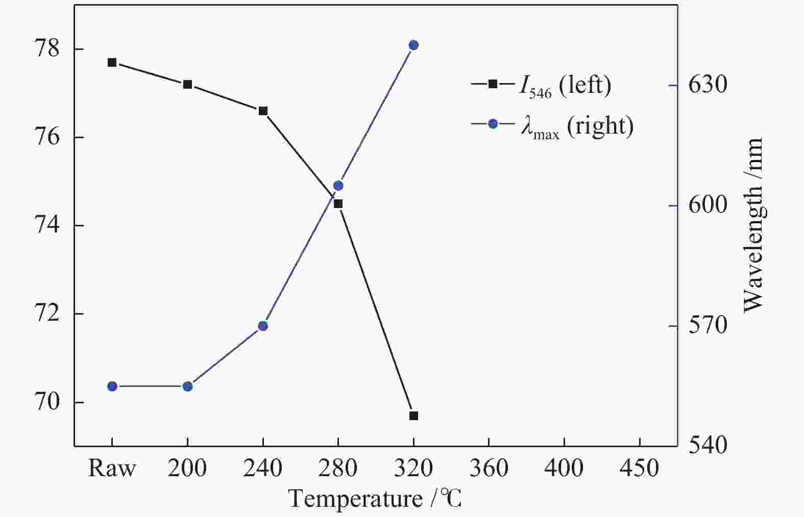
图 6 不同热转化温度下木栓质体的荧光特性
Figure 6 Fluorescence properties of suberinite at low temperature thermal conversion
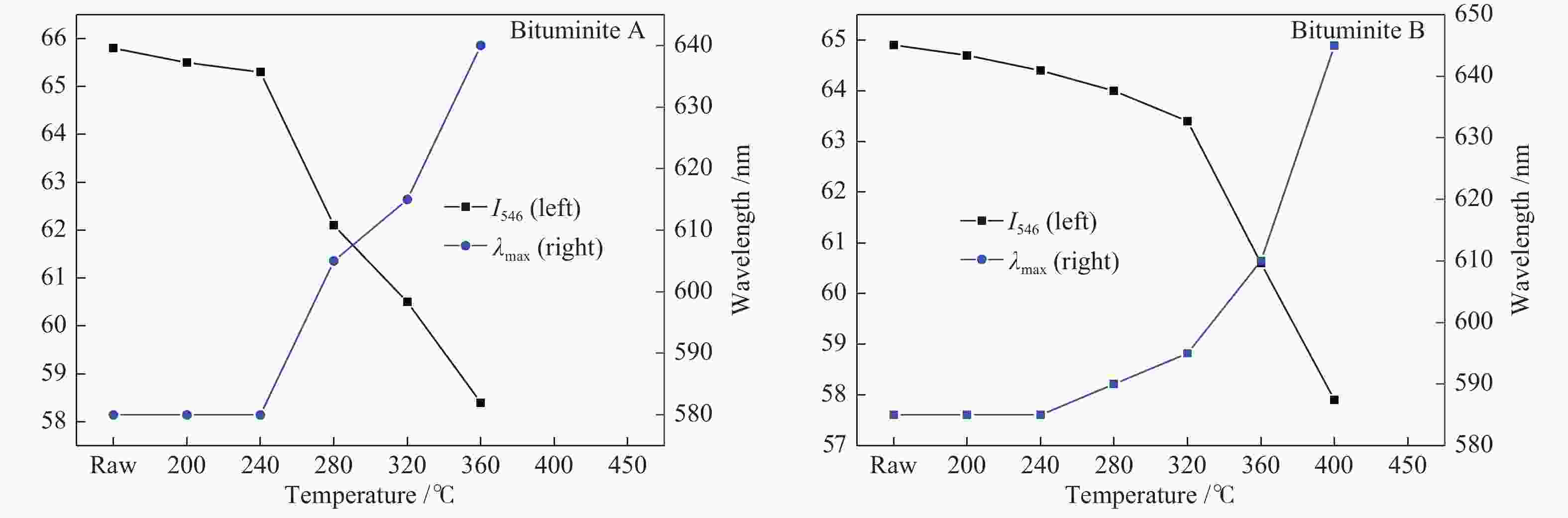
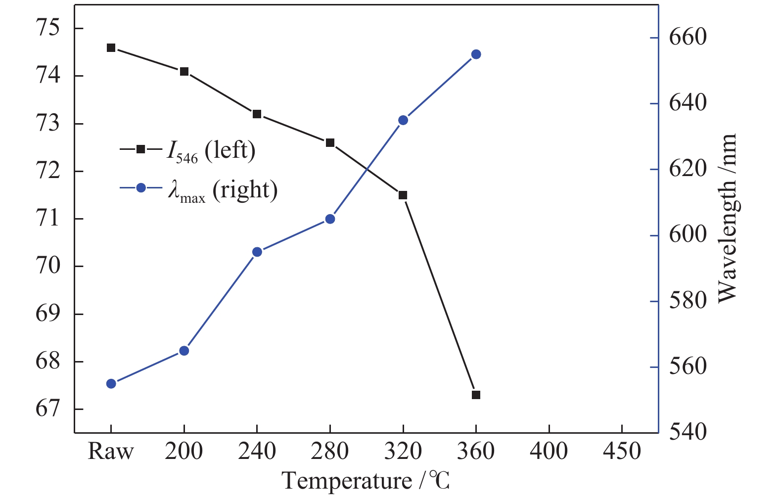
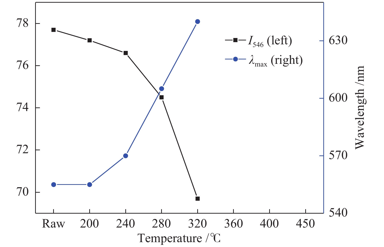
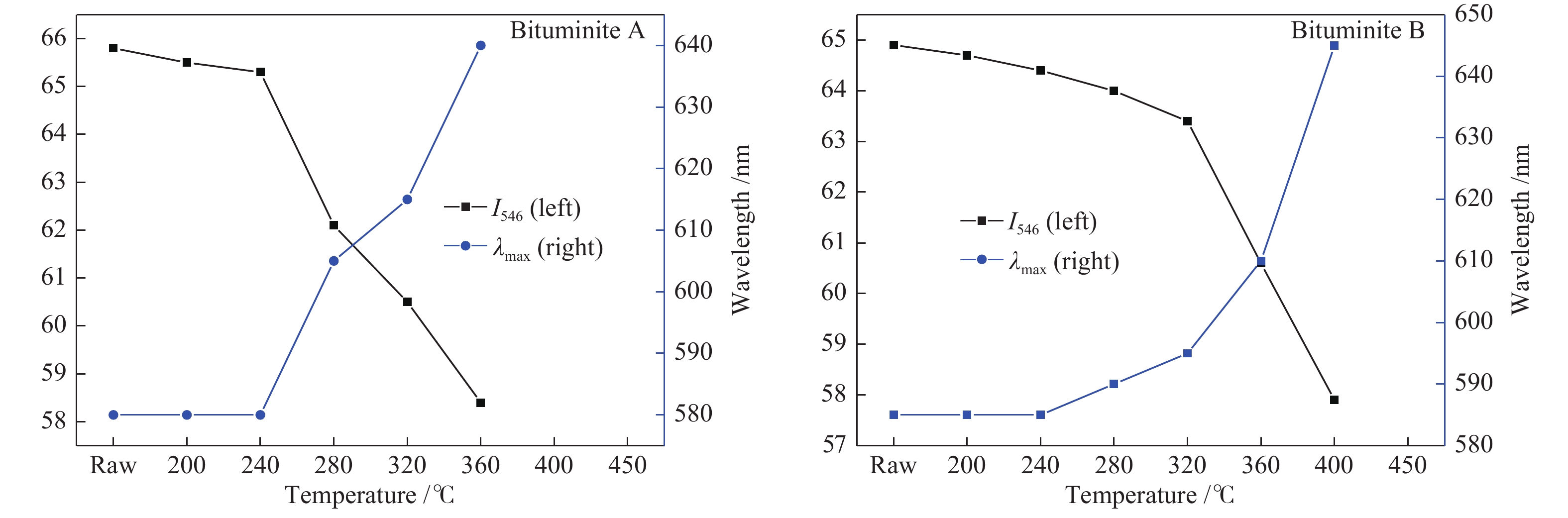
图 7 不同热转化温度下沥青质体的荧光特性
Figure 7 Fluorescence properties of bituminite at low temperature thermal conversion
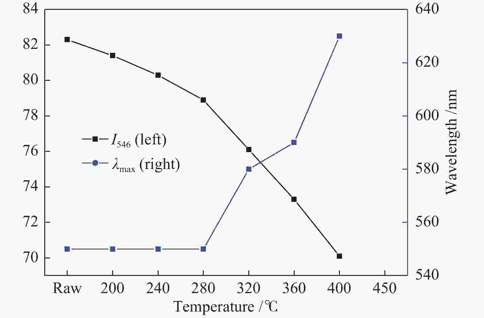
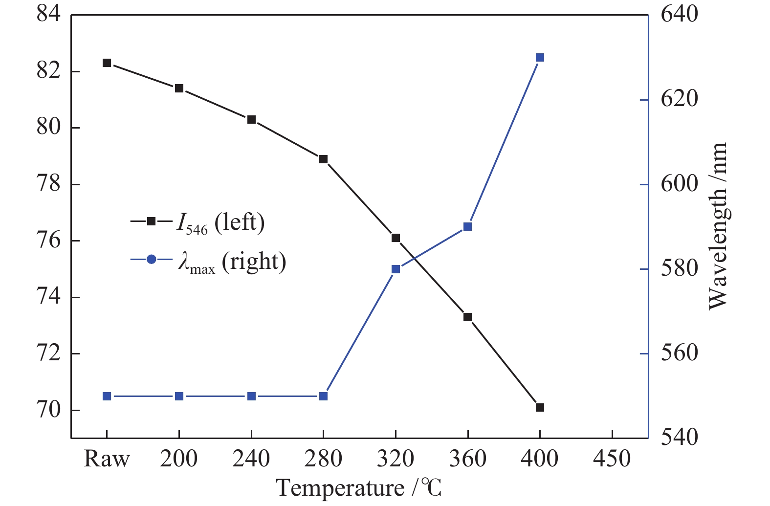
图 8 不同热转化温度下藻类体的荧光特性
Figure 8 Fluorescence properties of alginite at low temperature thermal conversion
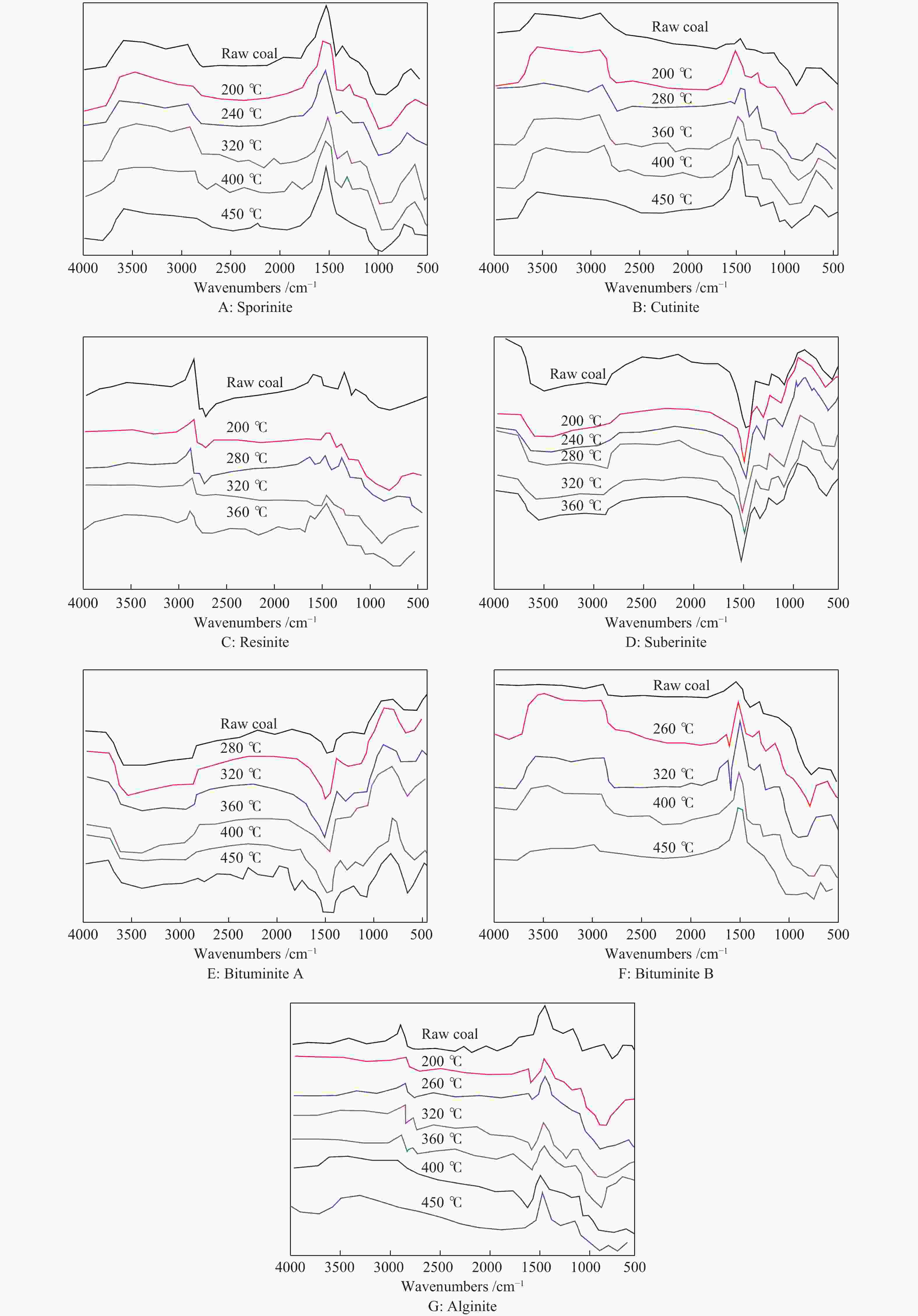
图 9 不同温度下各种壳质组分的Micro-FTIR谱图
Figure 9 Micro-FTIR spectrogram of typical liptinite at low temperature thermal conversion

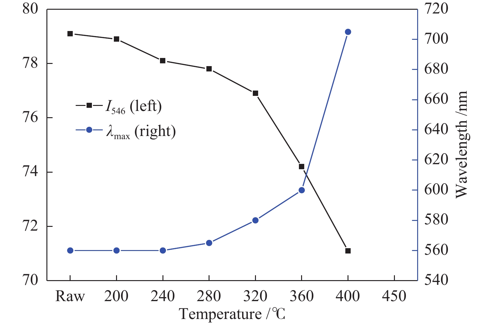
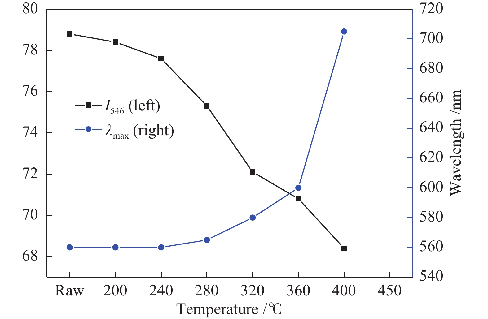
图 10 煤中典型壳质组的Micro-FTIR光谱特征参数随温度变化规律
Figure 10 Characteristic Micro-FTIR parameters of typical liptinite at low temperature thermal conversion
表 1 实验样品的基本煤质特征
Table 1 Basic characteristics of the sample
Proximate analysis w/% Ultimate analysis wdaf/% Mad Ad Vdaf FCd C H N S O* 2.57 4.05 38.20 59.29 82.22 5.05 0.94 0.39 11.40 St,d/% Calorific value/(MJ·kg−1) GR.I Gray-King assay Qgr,d Q net,ar waterad/% CRad/% tarad/% coke type loss/% 0.37 32.05 29.99 0 6.0 71.6 12.7 C 9.7 *:by difference  下载: 导出CSV
表 2 实验样品的煤岩特征
Table 2 Petrographic characteristics of the sample
Maceral group
(vol, mineral free)$ R_{\max }^ \circ /{\rm{\% }} $ Maceral in
liptinite groupvitrinite inertinite liptinite Sp Cu Re Sub Bt Alg 56.4 38.8 4.8 0.56 1.2 1.0 0.8 0.6 0.8 0.4 Sp: Sporinite; Cu: Cutinite; Re: Resinite; Sub: Subernite;
Bt: Bituminite; Alg: Alginite; $ R_{\max }^ \circ /{\rm{\% }} $: maximum reflectance of vitrinite
下载: 导出CSV
-
[1] 韩德馨. 中国煤岩学[M]. 徐州: 中国矿业大学出版社, 1996: 266−275.HAN De-xin. China Coal Petrology[M]. Xuzhou: China University of Mining and Technology Press, 1996: 266−275. [2] 陈鹏. 中国煤炭性质、分类和利用[M]. 北京: 化学工业出版社, 2007: 33−35.CHEN Peng. Nature, Classification and Utilization of Coal in China[M]. Beijing: Chemical Industry Press, 2007: 33−35. [3] 孙旭光, 陈建平, 郝多虎. 塔里木盆地煤显微组分显微傅里叶红外光谱特征及意义[J]. 北京大学学报(自然科学版),2001,37(6):832−838.SUN Xu-guang, CHEN Jian-ping, HAO Duo-hu. Micro-FTIR spectroscopy of macerals in coalsfrom the tarim basin[J]. Acta Sci Nat Univ Pekin,2001,37(6):832−838. [4] CHEN Y, MASTALERZ M, SCHIMMELMANN A. Characterization of chemical functional groups in macerals across different coal ranksvia micro-FTIR spectroscopy[J]. Int J Coal Geol,2012,104:22−33. doi: 10.1016/j.coal.2012.09.001 [5] 余晓露, 白帆, 李志明. 衰减全反射-显微傅立叶变换红外光谱原位分析煤有机显微组分[J]. 石油实验地质,2012,34(6):664−670.YU Xiao-lu, BAI Fan, LI Zhi-ming. Application of attenuated total reflectance-micro-Fourier transforminfrared (ATR-FTIR) spectroscopy to in situ study of coal macerals[J]. Pet Geol Exp,2012,34(6):664−670. [6] 刘大锰, 杨起, 汤达祯. 鄂尔多斯盆地煤显微组分的micro-FTIR研究[J]. 地球科学,1998,23(1):3−5.LIU Da-meng, YANG Qi, TANG Da-zhen. Micro-FTIR analysis of maceralsin coals from the ordos basin[J]. Earth Sci,1998,23(1):3−5. [7] 常海洲, 曾凡桂, 李文英, 李军, 贾建波, 谢克昌. 西北地区侏罗纪煤显微组分结构的Micro-FTIR研究[J]. 光谱学与光谱分析,2008,28(7):1535−1538. doi: 10.3964/j.issn.1000-0593.2008.07.022CHANG Hai-zhou, ZENG Fan-gui, LI Wen-ying, LI Ju, JIA Jian-bo, XIE Ke-chang. Micro-FTIR Study on structure of macerals from Jurassic coals in Northwestern China[J]. Spectro Spectral Anal,2008,28(7):1535−1538. doi: 10.3964/j.issn.1000-0593.2008.07.022 [8] MASTALERZ M, BUSTIN R M. Application of reflectance micro-Fourier Transforminfrared analysis to the study of coal macerals: An example from the Late Jurassic to Early Cretaceouscoals of the Mist Mountain Formation, British Columbia, Canada[J]. Int J Coal Geol,1996,32:55−67. doi: 10.1016/S0166-5162(96)00030-4 [9] LI Z S, FREDERICKS P M, RINTOUL L, WARD C R. Application of attenuated total reflectance micro-Fourier transforminfrared (ATR-FTIR) spectroscopy to the study of coal macerals: Examples from the Bowen Basin, Australia[J]. Int J Coal Geol,2007,70:87−94. doi: 10.1016/j.coal.2006.01.006 [10] MORGA R. Chemical structure of semifusinite and fusinite of steam and coking coal from theUpper Silesian Coal Basin (Poland) and its changes during heating as inferred frommicro-FTIR analysis[J]. Int J Coal Geol,2010,84:1−15. doi: 10.1016/j.coal.2010.07.003 [11] CHEN Y, CARO L D, MASTALERZ M, SCHIMMELMANN A, BLANDON A. Mapping the chemistry of resinite, funginite and associatedvitrinite in coal with micro-FTIR[J]. J Microsc,2013,249(1):69−81. doi: 10.1111/j.1365-2818.2012.03685.x [12] GUO Y, BUSTIN R. M. Micro-FTIR spectroscopy of liptinite macerals incoal[J]. Int J Coal Geol,1998,36:259−275. doi: 10.1016/S0166-5162(97)00044-X [13] GUO Y, RENTON J J, PENN J H. FTIR micro spectroscopy of particular liptinite-(lopinite-) rich, Late Permian coals from Southern China[J]. Int J Coal Geol,1996,29:187−197. doi: 10.1016/0166-5162(95)00024-0 [14] 余海洋, 孙旭光, 焦宗福. 华南晚二叠世“树皮体”显微傅里叶红外光谱(Micro-FTIR)特征及意义[J]. 北京大学学报(自然科学版),2004,40(6):879−885.YU Hai-yang, SUN Xu-guang, JIAO Zong-fu. Characteristics and implications of Micro- FTIR spectroscopy of Barkinite from Upper Permian Coals, South China[J]. Acta Sci Nat Univ Pekin,2004,40(6):879−885. [15] WANG S Q, LIU S M, SUN Y B, JIANG D, ZHANG X M. Investigation of coal components of Late Permian different ranks bark coal using AFM and Micro-FTIR[J]. Fuel,2017,187:51−57. doi: 10.1016/j.fuel.2016.09.049 [16] LYONS P C, OREM W H, MASTALERZ M, ZODROW E L, VIETHREDEMANN A, BUSTIN R M. 13C NMR, micro-FTIR and fluorescence spectra, andpyrolysis-gas chromatograms of coalified foliage oflate Carboniferous medullosan seed ferns, NovaScotia, Canada: Implications for coalification and chemotaxonomy[J]. Int J Coal Geol,1995,27:227−248. doi: 10.1016/0166-5162(94)00024-T [17] MASTALERZ M, BUSTIN R M. Electron microprobe and micro-FTIR analysesapplied to maceral chemistry[J]. Int J Coal Geol,1993,24:333−345. doi: 10.1016/0166-5162(93)90018-6 [18] 姚素平, 张景荣, 金奎励. 用显微荧光和显微傅立叶红外光谱研究显微组分的热演化规律[J]. 沉积学报,1996,14(3):3−12.YAO Su-ping, ZHANG Jing-rong, JIN Kui-li. Studying individual macerals using FTIR microspectroscopy and fluorescence spectroscopy on the thermal evolution[J]. Acta Sedimentol Sin,1996,14(3):3−12. [19] 李东涛, 李文, 孙庆雷, 李保庆. 原位漫反射红外光谱中采用新的实验手段研究煤岩显微组分中的氢键[J]. 高等学校化学学报,2003,24(4):703−706. doi: 10.3321/j.issn:0251-0790.2003.04.027LI Dong-tao, LI Wen, SUN Qing-lei, LI Bao-qing. A study on hydrogen bond in coal macerals with in situ diffusereflectance FTIR by using a new experimental method[J]. Chem Chin Univ,2003,24(4):703−706. doi: 10.3321/j.issn:0251-0790.2003.04.027 [20] 周师庸. 应用煤岩学[M]. 北京: 冶金工业出版社, 1985: 77−85.ZHOU Shi-yong. Applied Coal Petrology[M]. Beijing: Metallurgical Industry Press, 1985: 77−85. [21] 周炎如. 应用显微FT-IR光谱技术“原位”研究沉积岩中生油母质──干酪根[J]. 沉积学报,1994,12(4):22−30.ZHOU Yan-ru. Situ-study on generative Kerogen in sedimentary rock by FTIR- microspectrometry technique[J]. Acta Sedimentol Sin,1994,12(4):22−30. [22] JOHNSTON M N, HOWER J C, RICH F J. Notes on the origin of the resinite-rich “pine needle” lithotype of the Cretaceous Cambria coal, Weston County, Wyoming[J]. Int J Coal Geol,2014,130:66−69. doi: 10.1016/j.coal.2014.05.008 [23] 王飞宇, 何萍, 赵长毅. 新疆侏罗纪煤中木栓质体的特征和演化[J]. 煤田地质与勘探,1995,(6):18−24.WANG Fei-yu, HE ping, ZHAO Chang-yi. Suberinite in Jurassic coals of xinjiang[J]. Coal Geol Explor,1995,(6):18−24. [24] 王越, 高燕, 白向飞, 武琳琳. 桦甸油页岩有机岩相特征及其富集特性[J]. 燃料化学学报,2016,44(3):321−327. doi: 10.3969/j.issn.0253-2409.2016.03.009WANG Yue, GAO Yan, BAI Xiang-fei, WU Lin-lin. Petrology and enrichment characteristics of organic matters in Huadian oil shale[J]. J Fuel Chem Technol,2016,44(3):321−327. doi: 10.3969/j.issn.0253-2409.2016.03.009 [25] PICKEL W, KUS J, FLORES D, KALAITZIDIS S, CHRISTANIS K, CARDOTT BJ, MISZ-KENNAN M, RODRIGUES S, HENTSCHEL A, HAMOR-VIDO M, CROSDALE P, WAGNER N, ICCP. Classification of liptinite-ICCP System 1994[J]. Int J Coal Geol,2017,169:40−61. doi: 10.1016/j.coal.2016.11.004 [26] 金奎励. 当代煤及有机岩研究新技术[M]. 北京: 地质出版社, 1997: 55−73.JIN Kui-li. New Technology of Coal and Organic Petrology[M]. Beijing: Geological Publishing House, 1997: 55−73. [27] LIN R, DAVIS A. A fluorogeochemical model for coal macerals[J]. Org Geochem,1988,12(4):363−374. doi: 10.1016/0146-6380(88)90010-1 [28] 韩志文, 周怡. 煤和油源岩的荧光性研究[M]. 北京: 地质出版社, 1993: 1−4.HAN Zhi-wen, ZHOU Yi. Fluorescence Characteristics of Coal and Oil-source Rocks[M]. Beijing: Geological Publishing House, 1993: 1−4. [29] GANZ H, KALKREUTH W. Application of infrared spectroscopy to the classificationof kerogen-types and the evaluation of source rock and oil shale[J]. Fuel,1987,66:708−711. doi: 10.1016/0016-2361(87)90285-7 [30] 斯塔赫. 斯塔赫煤岩学教程[M]. 北京: 煤炭工业出版社, 1990: 184−201.STACH E. Stach’s Textbook of Coal Petrology[M]. Beijing: Coal Industry Press, 1990: 184−201. [31] PRADIER B, LANDAIS P, RPCHDI A, DAVIS A. Chemical basis of fluorescence alteration of crude oils and kerogens-Ⅱ: Fluorescence and infrared micro-spectrometric analysis of vitrinite and liptinite[J]. Org Geochem,1992,18(3):241−248. doi: 10.1016/0146-6380(92)90065-6 [32] WANG S Q, TANG Y G, SCHOBERT H H, JIANG D, SUN Y B, GUO Y N, SU Y F, YANG S P. Application and thermal properties of hydrogen-rich bark coal[J]. Fuel,2015,162:121−127. -

 点击查看大图
计量
- 文章访问数: 250
- HTML全文浏览量: 55
- PDF下载量: 20
- 被引次数: 0
